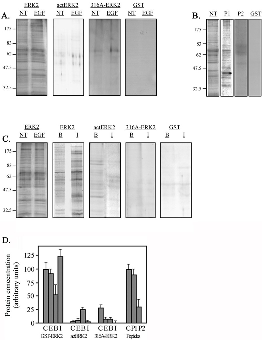
Fig. 1. Detection of ERK2-interacting proteins by silver staining. A. Extracts of quiescent or EGF-stimulated (50 ng/ml, 15 min) Rat1 cells (3 mg protein) were loaded on 0.5 ml columns of GST-ERK2, GST-actERK2, GST-316A-ERK2 and GST alone and developed as described. The 0.2M NaCl eluates were subjected to SDS-PAGE and silver stained. B. Rat1 extracts from non-stimulated cells were loaded on the GST-ERK2 columns in the presence of a peptide (0.5 mg/ml) derived from FAR1 (P1), from the CRS of ERK2 (P2) or without peptide and the eluates were processed as above. C. Extracts (0.5 mg protein) from BAPTA-AM (15 µM, 60 min) and ionomycin (1 µM, 30 min) were loaded on the indicated columns, which were processed in the presence of EGTA (1.5 mM, for BAPTA-AM extracts) or CaCl2 (1 mM, for ionomycin column), and processed as above. The eluates of non-treated and EGF stimulated cells is brought for comparison. D. The protein concentrations in the various eluates were determined by Bradford protein assay. The barographs represent averages of three distinct experiments.